
Anatomy and function
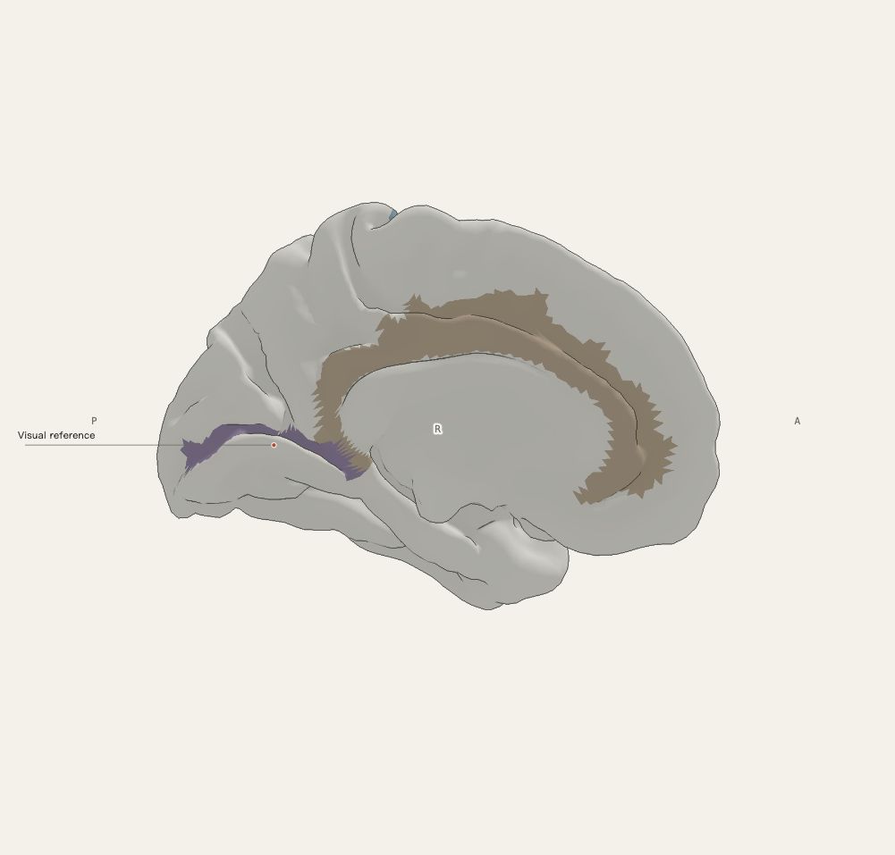
Primary visual cortex lies mainly along the banks of the calcarine sulcus on the medial surface of the occipital lobe. Visual information also reaches multiple extrastriate regions involved in shape, colour and motion processing.
Where to look in the model
The left and right calcarine sulcus parcels provide a medial visual-location reference. Use the medial view.
What this view does not establish
The highlight is a sulcal anatomical parcel, not the full extent of V1. Retinotopic organisation is not shown.
Open in 3DSources and precision
Cortex uses the FreeSurfer fsaverage5 template surface, with 10,242 vertices per hemisphere. Functional references use selected Destrieux sulcal and gyral parcels. Deep structures use fsaverage aseg segmentations. The Sereno2020 cerebellar research surface preserves folial geometry, but comes from another specimen; its assembled placement is schematic, without medical registration across templates. Arterial paths are manually drawn teaching schematics, not CTA/MRA reconstructions.
An educational schematic prototype for understanding spatial relationships. Item-by-item independent neuroanatomical review is pending. Functional highlights are not results of an individual examination and do not support individual diagnosis or treatment decisions.