
Anatomy and function
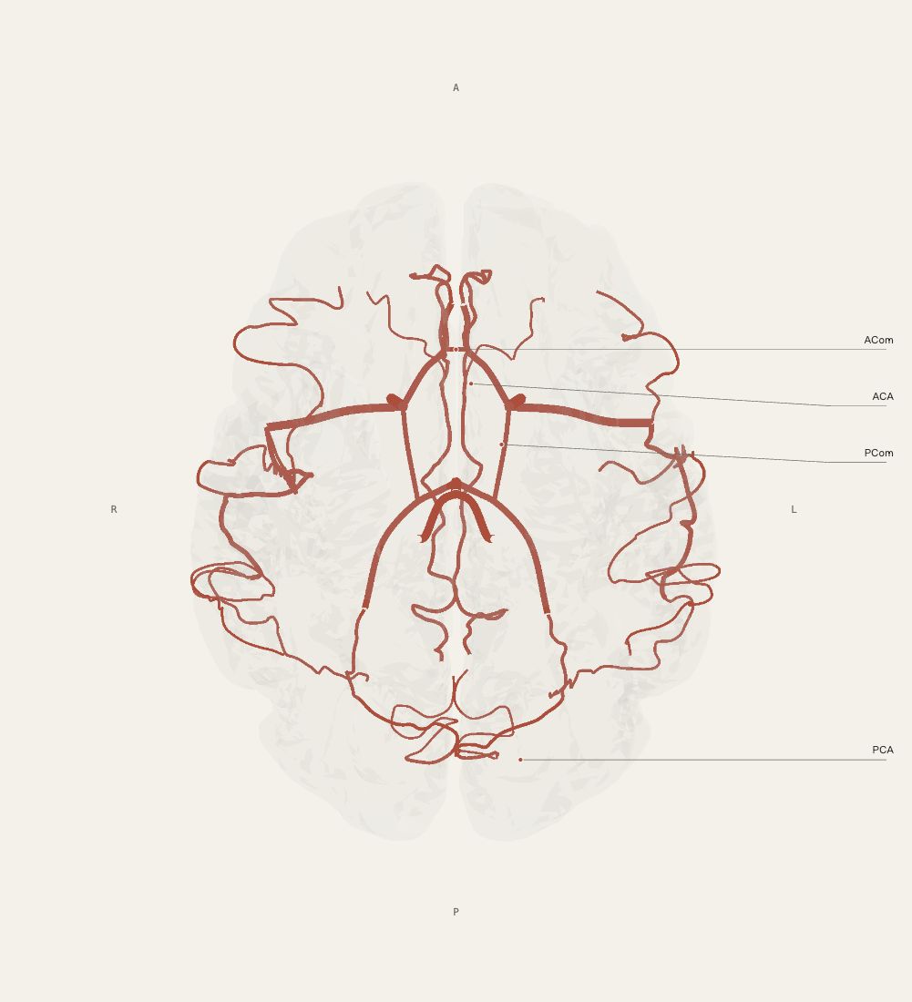
The anterior communicating artery connects the two anterior cerebral arteries. The posterior communicating arteries usually connect each internal carotid artery with the posterior cerebral artery on the same side. These connections help illustrate relationships between the anterior and posterior circulations.
Where to look in the model
The inferior view shows schematic connections between the terminal internal carotid arteries, ACAs, PCAs and communicating arteries, together with some input vessels and branches.
What this view does not establish
The circle of Willis is not separately or precisely segmented in this model. The ACA and PCA drawings extend beyond the ring; the MCA and basilar artery are not part of the ring itself. Connections are idealised and do not represent individual variants or collateral capacity.
Open in 3DSources and precision
Cortex uses the FreeSurfer fsaverage5 template surface, with 10,242 vertices per hemisphere. Functional references use selected Destrieux sulcal and gyral parcels. Deep structures use fsaverage aseg segmentations. The Sereno2020 cerebellar research surface preserves folial geometry, but comes from another specimen; its assembled placement is schematic, without medical registration across templates. Arterial paths are manually drawn teaching schematics, not CTA/MRA reconstructions.
An educational schematic prototype for understanding spatial relationships. Item-by-item independent neuroanatomical review is pending. Functional highlights are not results of an individual examination and do not support individual diagnosis or treatment decisions.